Diagrams
Figures you can use
Every diagram on this site, drawn from the cited literature rather than traced from a stock illustration. Each one is free to put on your own page, including commercially, as long as a visible link points back here. They follow your reader's light or dark theme.
Why we give them away: the good renderings of these — the Norwood scale especially — belong to clinics who use them to collect phone numbers. There should be a free, accurate one.
Laboratory normal against the hair-relevant range#
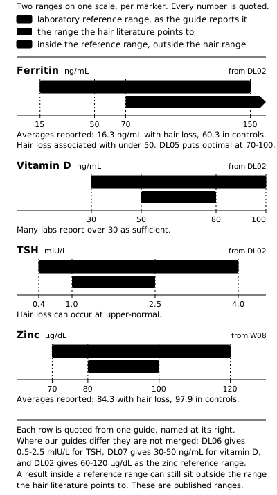
Laboratory reference ranges against the ranges the hair literature points to, for ferritin, vitamin D, TSH and zinc
Four blood markers, each drawn on its own scale as two bars: the laboratory reference range, and the narrower range the hair literature points to. Ferritin, in nanograms per millilitre: the laboratory reference range for women runs 15 to 150, while the guide gives 70 and above as optimal for hair, so the stretch from 15 to 70 is inside the reference range and outside the hair range; hair loss is associated with levels under 50, and reported averages are 16.3 in hair loss patients against 60.3 in controls, both of them inside the reference range. That 15 to 150 is the range the guide gives for women. Vitamin D, in nanograms per millilitre: reference range 30 to 100, hair range 50 to 80, with shaded stretches from 30 to 50 below the hair range and from 80 to 100 above it; many laboratories report over 30 as sufficient. TSH, in milli-international units per litre: reference range 0.4 to 4.0, hair range 1.0 to 2.5; here one shaded stretch sits above the hair range, from 2.5 to 4.0, because hair loss can occur at upper-normal, and a second runs from 0.4 to 1.0 below it. Zinc, in micrograms per decilitre: reference range 70 to 120, hair range 80 to 100, with shaded stretches from 70 to 80 and from 100 to 120; reported averages are 84.3 in hair loss patients against 97.9 in controls. Ferritin, vitamin D and TSH are quoted from the blood test guide DL02, and zinc from the women’s blood test guide W08. Our guides do not always agree and the ranges are not merged: DL06 gives 0.5 to 2.5 for TSH, DL07 gives 30 to 50 nanograms per millilitre for vitamin D, and DL02 gives 60 to 120 micrograms per decilitre as the zinc reference range. A result inside a reference range can still sit outside the range the hair literature points to.
Two ranges on one scale, per marker. Every number is quoted.
laboratory reference range, as the guide reports it the range the hair literature points to inside the reference range, outside the hair range
Ferritinng/mL
from DL02
lab reference (women)
hair literature
15 50 70 150
Averages reported: 16.3 ng/mL with hair loss, 60.3 in controls. Hair loss associated with under 50. DL05 puts optimal at 70-100.
Vitamin Dng/mL
from DL02
lab reference
hair literature
30 50 80 100
Many labs report over 30 as sufficient.
TSHmIU/L
from DL02
lab reference
hair literature
0.4 1.0 2.5 4.0
Hair loss can occur at upper-normal.
Zincµg/dL
from W08
lab reference
hair literature
70 80 100 120
Averages reported: 84.3 with hair loss, 97.9 in controls.
Four markers, each drawn twice on one scale: the laboratory reference range the guides report, and the narrower range the hair literature points to. The shaded stretch of each reference bar is where a result can be reported as normal and still sit outside the hair range. Each row is quoted from a single guide, named on the row; where our guides disagree the ranges are printed, not merged. Appears in Hair Loss Blood Tests: What to Request and Why (2026) · Ferritin and Hair Loss: Optimal Levels for Hair Growth (2026) .
Use this figure on your own site
Free to use anywhere, including commercially, if you keep a visible link back to this page. It is drawn from the sources cited beneath it and follows your reader's light or dark theme.
Open the SVG directly · All figures
The DHT pathway, and where each drug blocks it#
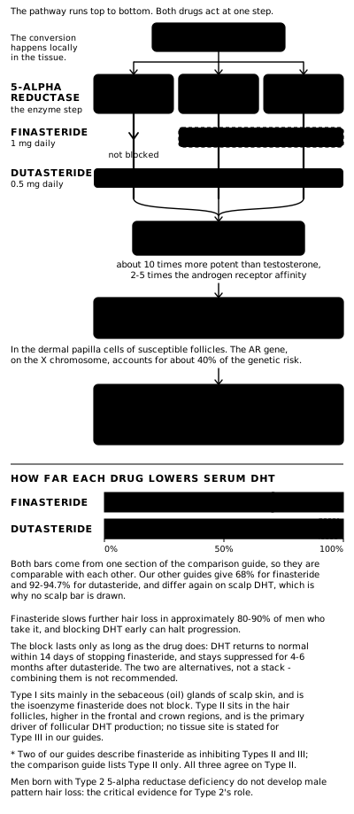
The androgen pathway that drives pattern hair loss, and the step finasteride and dutasteride block
A flow chart read top to bottom. Testosterone at the top feeds into one enzyme step, 5-alpha reductase, drawn as three parallel channels: Type I, sited in the sebaceous or oil glands of scalp skin; Type II, sited in the hair follicles, higher in the frontal and crown regions and the primary driver of follicular DHT production; and Type III, for which our guides state no tissue site. Two horizontal bars cross the channels. The finasteride bar, at 1 mg daily, closes Type II solidly and Type III in a lighter shade marked with an asterisk, and leaves the Type I channel open with an arrow showing the pathway continuing past it. The dutasteride bar, at 0.5 mg daily, closes all three channels. The channels then converge into DHT, dihydrotestosterone, described as about 10 times more potent than testosterone with 2 to 5 times the androgen receptor affinity. An arrow leads to the androgen receptor, drawn as a cup with DHT seated in it, in a genetically susceptible follicle: in the dermal papilla cells, with the AR gene on the X chromosome accounting for about 40 percent of genetic risk. The last arrow leads to miniaturisation: anagen shortens, telogen lengthens, and each cycle produces a thinner, shorter hair until terminal hair becomes vellus. Below the pathway, two bars compare serum DHT reduction on a 0 to 100 percent scale: finasteride 70 to 71 percent, dutasteride 90 to 98 percent. Both bars come from one section of the comparison guide so they are comparable with each other; our other guides give 68 percent for finasteride and 92 to 94.7 percent for dutasteride, and differ again on scalp DHT, which is why no scalp bar is drawn. Notes record that finasteride slows further hair loss in approximately 80 to 90 percent of men who take it and that blocking DHT early can halt progression; that the block lasts only as long as the drug does, with DHT returning to normal within 14 days of stopping finasteride and staying suppressed for 4 to 6 months after dutasteride, the two being alternatives rather than a stack; that our guides disagree about whether finasteride reaches Type III; and that men born with Type 2 5-alpha reductase deficiency do not develop male pattern hair loss.
The pathway runs top to bottom. Both drugs act at one step. TESTOSTERONE The conversion happens locally in the tissue. 5-ALPHA REDUCTASE the enzyme step TYPE I oil glands TYPE II hair follicles TYPE III site not stated BLOCKS BLOCKS * FINASTERIDE 1 mg daily not blocked BLOCKS BLOCKS BLOCKS DUTASTERIDE 0.5 mg daily DHT dihydrotestosterone about 10 times more potent than testosterone, 2-5 times the androgen receptor affinity ANDROGEN RECEPTOR in a genetically susceptible follicle In the dermal papilla cells of susceptible follicles. The AR gene, on the X chromosome, accounts for about 40% of the genetic risk. MINIATURISATION Anagen shortens, telogen lengthens, and each successive cycle produces a thinner, shorter hair: terminal hairs become vellus. HOW FAR EACH DRUG LOWERS SERUM DHT 70-71% FINASTERIDE 90-98% DUTASTERIDE 0% 50% 100% Both bars come from one section of the comparison guide, so they are comparable with each other. Our other guides give 68% for finasteride and 92-94.7% for dutasteride, and differ again on scalp DHT, which is why no scalp bar is drawn. Finasteride slows further hair loss in approximately 80-90% of men who take it, and blocking DHT early can halt progression. The block lasts only as long as the drug does: DHT returns to normal within 14 days of stopping finasteride, and stays suppressed for 4-6 months after dutasteride. The two are alternatives, not a stack - combining them is not recommended. Type I sits mainly in the sebaceous (oil) glands of scalp skin, and is the isoenzyme finasteride does not block. Type II sits in the hair follicles, higher in the frontal and crown regions, and is the primary driver of follicular DHT production; no tissue site is stated for Type III in our guides. * Two of our guides describe finasteride as inhibiting Types II and III; the comparison guide lists Type II only. All three agree on Type II. Men born with Type 2 5-alpha reductase deficiency do not develop male pattern hair loss: the critical evidence for Type 2's role.
Testosterone becomes DHT at a single enzyme step with three isoenzymes; DHT then binds the androgen receptor in a genetically susceptible follicle, and the follicle miniaturises. Finasteride closes Type II, dutasteride closes all three, which is why lowering DHT slows the loss and why the two drugs lower it by different amounts. Appears in DHT and Hair Loss: Complete Explanation · Complete Guide to Finasteride for Hair Loss · Dutasteride vs Finasteride: Which Is Better for Hair Loss? .
Use this figure on your own site
Free to use anywhere, including commercially, if you keep a visible link back to this page. It is drawn from the sources cited beneath it and follows your reader's light or dark theme.
Open the SVG directly · All figures
FUE and FUT: what each one takes, and what each one leaves#
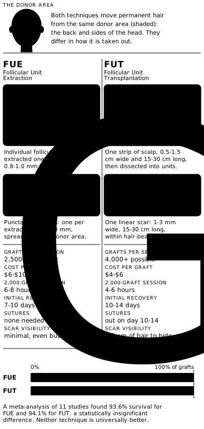
FUE and FUT compared: what each technique removes from the donor area, and what scar it leaves
At the top, a head seen from behind, with a shaded horseshoe band across the back and up both sides: the donor area both techniques harvest from. Below it, two columns. Left column, FUE, Follicular Unit Extraction: a patch of hair-bearing donor scalp is shown perforated by many small round holes, and beneath it four follicular units, each drawn inside the dotted ring of the punch that cut it out. Individual follicular units are extracted one by one with punches of 0.8 to 1.0 millimetres, with no scalpel incision. The healed panel below shows a dot scar at every one of those same sites, scattered across the donor area, each 0.8 to 1.0 millimetres across, minimal even at buzz-cut length. FUE takes 2,500 to 3,000 grafts at most per session, costs 6 to 10 dollars a graft, takes 6 to 8 hours for 2,000 grafts, needs 7 to 10 days of initial recovery, and needs no sutures. Right column, FUT, Follicular Unit Transplantation: the same patch of donor scalp with a single strip missing from the middle, drawn as a dashed gap, and beneath it that strip shown divided into the individual follicular units it is dissected into under microscopes. The strip is 0.5 to 1.5 centimetres wide and 15 to 30 centimetres long. The healed panel below shows one linear scar where the two edges were closed, 1 to 3 millimetres wide and 15 to 30 centimetres long, concealed within hair-bearing scalp, with a few hairs drawn growing through it as trichophytic closure allows; 2 to 3 centimetres of hair is the minimum needed to hide it. FUT allows 4,000 or more grafts per session, costs 4 to 6 dollars a graft, takes 4 to 6 hours for 2,000 grafts, needs 10 to 14 days of initial recovery, and sutures come out on day 10 to 14. At the foot, two bars on a full zero to one hundred percent scale show mean graft survival: 93.6 percent for FUE and 94.1 percent for FUT. A meta-analysis of 11 studies found that difference statistically insignificant, and neither technique is universally better.
THE DONOR AREA
Both techniques move permanent hair from the same donor area (shaded): the back and sides of the head. They differ in how it is taken out.
FUE
Follicular Unit Extraction
WHAT IS TAKEN
Individual follicular units extracted one by one with 0.8-1.0 mm punches.
WHAT IS LEFT
Punctate dot scars: one per extraction, 0.8-1.0 mm, spread over the donor area.
GRAFTS PER SESSION 2,500-3,000 max COST PER GRAFT $6-$10 2,000-GRAFT SESSION 6-8 hours INITIAL RECOVERY 7-10 days SUTURES none needed SCAR VISIBILITY minimal, even buzz cut
FUT
Follicular Unit Transplantation
WHAT IS TAKEN
One strip of scalp, 0.5-1.5 cm wide and 15-30 cm long, then dissected into units.
WHAT IS LEFT
One linear scar: 1-3 mm wide, 15-30 cm long, within hair-bearing scalp.
GRAFTS PER SESSION 4,000+ possible COST PER GRAFT $4-$6 2,000-GRAFT SESSION 4-6 hours INITIAL RECOVERY 10-14 days SUTURES out on day 10-14 SCAR VISIBILITY 2-3 cm of hair to hide
Graft survival: no meaningful difference
0%
100% of grafts
FUE
93.6%
FUT
94.1%
A meta-analysis of 11 studies found 93.6% survival for
FUE and 94.1% for FUT: a statistically insignificant
difference. Neither technique is universally better.
Both techniques move hair from the same donor area. FUT removes a single strip of scalp and dissects it into follicular units, leaving one linear scar; FUE punches units out individually, leaving scattered dot scars. Published graft survival is 93.6% for FUE and 94.1% for FUT, a difference the meta-analysis calls statistically insignificant. Appears in Hair Transplant FUE vs FUT: Complete Comparison (2026) .
Use this figure on your own site
Free to use anywhere, including commercially, if you keep a visible link back to this page. It is drawn from the sources cited beneath it and follows your reader's light or dark theme.
Open the SVG directly · All figures
The hair growth cycle#
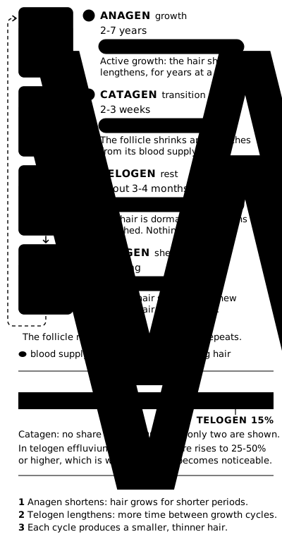
The four phases of the hair growth cycle, with their published durations and shares
A follicle is drawn four times in sequence. 1. Anagen, the growth phase, lasts 2 to 7 years for scalp hair; the shaft lengthens and the follicle sits deep against its blood supply; about 85 percent of scalp hair is in anagen at any time. 2. Catagen, the transition phase, lasts 2 to 3 weeks; the follicle shrinks and detaches from its blood supply; it is a very brief transitional period and our sources give no share for it. 3. Telogen, the resting phase, lasts about 3 to 4 months; the hair is dormant but remains attached, held by its root while the blood supply sits detached below; about 15 percent of scalp hair is in telogen. 4. Exogen, shedding, is ongoing rather than timed; the old hair sheds as the new anagen hair pushes it out, which accounts for the 50 to 100 hairs a day shed in normal cycling. An arrow returns from exogen to anagen: the cycle repeats. A bar shows the anagen and telogen split, about 85 percent against about 15 percent; catagen is not on it, because no share is published for it. In telogen effluvium the resting share rises to 25 to 50 percent or higher. In pattern hair loss the cycle itself changes: anagen shortens, telogen lengthens, and each cycle produces a smaller, thinner hair.
1
ANAGENgrowth
2-7 years
about 85% of scalp hair
Active growth: the hair shaft
lengthens, for years at a time.
2
CATAGENtransition
2-3 weeks
very brief transition
The follicle shrinks and detaches
from its blood supply.
3
TELOGENrest
about 3-4 months
about 15% of scalp hair
The hair is dormant but remains
attached. Nothing is growing.
4
EXOGENshed
ongoing
50-100 hairs a day
The old hair sheds as the new
anagen hair pushes it out.
blood supply
root of the shedding hair
The follicle re-enters anagen: the cycle repeats.
Where scalp hair sits at any one moment
ANAGEN 85%
TELOGEN 15%
Catagen: no share in our sources, so only two are shown.
In telogen effluvium the resting share rises to 25-50%
or higher, which is when shedding becomes noticeable.
What pattern hair loss changes about the cycle
1 Anagen shortens: hair grows for shorter periods.2 Telogen lengthens: more time between growth cycles.3 Each cycle produces a smaller, thinner hair. One follicle through anagen, catagen, telogen and exogen, with the published duration of each phase and the share of scalp hair sitting in each at any one moment. Shedding is the end of a cycle, not a failure of one. Appears in Androgenetic Alopecia: Complete Guide · Telogen Effluvium: Causes and Recovery Timeline · How Long Until Hair Loss Treatment Works? The Complete Timeline .
Use this figure on your own site
Free to use anywhere, including commercially, if you keep a visible link back to this page. It is drawn from the sources cited beneath it and follows your reader's light or dark theme.
Open the SVG directly · All figures
The Ludwig scale, grades I to III#
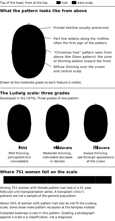
The Ludwig scale: grades I to III of female pattern hair loss, seen from above
Four diagrams of the top of a head, front at the top, with no face drawn. Solid tone is hair; pale slivers are scalp showing through between the hairs. A large reference head is drawn at the moderate grade and labelled: the band of hair at the frontal hairline is usually preserved; the part line widens along the midline, often the first sign of the pattern; the thinning zone is a triangle that widens toward the front, the shape called the Christmas tree or Olsen pattern; and the thinning is diffuse over the crown and central scalp. Below it the three Ludwig grades are drawn in a row. Grade I, mild: the part is barely widened and the thinning is concealable. Grade II, moderate: a noticeable decrease in density, with scalp visible either side of a wider part. Grade III, severe: the gaps run together and the scalp shows through, a see-through appearance at the crown. The hairs still standing are drawn finer at each grade because the mechanism is miniaturisation, not simple loss. In all three the front band of hair is unbroken. A bar shows the grades of 751 women in a 31-year follicular unit transplantation series: 40 percent Ludwig I, 45 percent Ludwig II, 15 percent Ludwig III, which is a surgical clinic's patients rather than the general population. Around 30 percent of women with pattern hair loss do not fit this scale; some show male-pattern recession at the temples instead. Complete baldness is rare in this pattern.
Top of the head, front at the top.
hair
bare scalp
What the pattern looks like from above
Frontal hairline usually preserved Part line widens along the midline, often the first sign of the pattern “Christmas tree” pattern seen from above (the Olsen pattern): the zone of thinning widens toward the front Diffuse thinning over the crown and central scalp
Drawn at the moderate grade so each feature is visible.
The Ludwig scale: three grades
Developed in the 1970s. Three grades of one pattern.
I Mild Mild thinning, perceptible but concealable
II Moderate Moderate thinning, noticeable decrease in density
III Severe Severe thinning, see-through appearance at the crown
Where 751 women fell on the scale
I 40% II 45% III 15%
Among 751 women with female pattern hair loss in a 31-year
follicular unit transplantation series. A transplant clinic's
patients are not a sample of the general population.
About 30% of women with pattern hair loss do not fit the Ludwig
scale; some show male-pattern recession at the temples instead.
Complete baldness is rare in this pattern. Grading a photograph
against a scale is a classification, not a diagnosis.
Female pattern hair loss seen from above. The centre part widens into a zone that broadens toward the front — the "Christmas tree" pattern — while the band of hair at the frontal hairline stays. Grades I, II and III are the same pattern at increasing severity: hair thins and scalp shows through it, rather than an area going bald. Appears in Women's Hair Loss: The Complete Guide · Female Pattern Hair Loss: Treatment Options That Work .
Use this figure on your own site
Free to use anywhere, including commercially, if you keep a visible link back to this page. It is drawn from the sources cited beneath it and follows your reader's light or dark theme.
Open the SVG directly · All figures
How a follicle miniaturises#
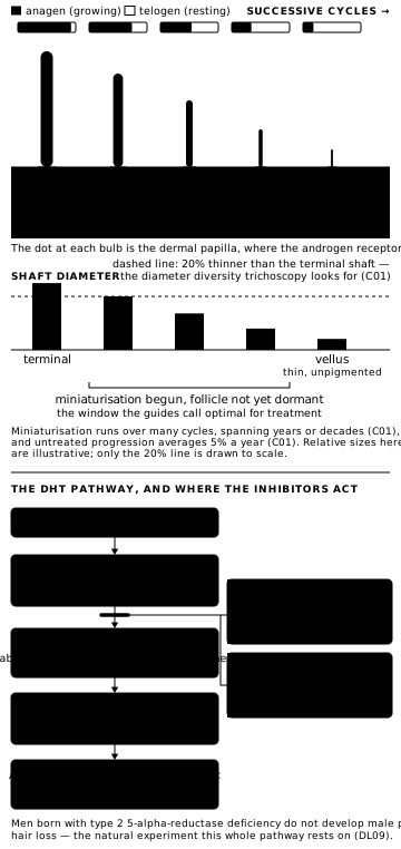
How a hair follicle miniaturises over successive cycles under DHT, and the DHT pathway and where 5-alpha-reductase inhibitors act
A row of five follicles reading left to right as one follicle at successive points in the process. Above each, a bar shows the cycle: the filled growing (anagen) phase shrinks and the outlined resting (telogen) phase grows. The hair shaft above the scalp gets shorter, finer and paler at each step, and the follicle beneath the skin gets shallower, ending as a barely visible vellus hair. Below, each shaft width is redrawn as a column on one shared scale, with a dashed line 20 per cent below the terminal shaft: hair diameter diversity above that 20 per cent is what trichoscopy looks for. A bracket marks the middle of the sequence as the window the guides emphasise, where miniaturisation has begun but the follicle is not yet dormant. Relative sizes are illustrative; only the 20 per cent line is drawn to scale. A downward chain: testosterone, then 5-alpha-reductase (type II in hair follicles, type I in sebaceous glands), then DHT — about 10 times more potent than testosterone with 2 to 5 times the receptor affinity — then the androgen receptor in the dermal papilla, whose gene carries about 40 per cent of genetic risk, and finally the outcome: a shorter growing phase and a thinner hair each cycle. A stop bar sits on the arrow from the enzyme to DHT, connected to two notes: finasteride 1 mg blocks type II and type III and lowers scalp DHT by 64 per cent and serum DHT by 68 per cent; dutasteride blocks types 1, 2 and 3 and lowers serum DHT by 92 to 94.7 per cent and scalp DHT by 51 per cent.
anagen (growing) telogen (resting) SUCCESSIVE CYCLES → SKIN The dot at each bulb is the dermal papilla, where the androgen receptors sit. SHAFT DIAMETER dashed line: 20% thinner than the terminal shaft — the diameter diversity trichoscopy looks for (C01) terminal vellus thin, unpigmented miniaturisation begun, follicle not yet dormant the window the guides call optimal for treatment Miniaturisation runs over many cycles, spanning years or decades (C01), and untreated progression averages 5% a year (C01). Relative sizes here are illustrative; only the 20% line is drawn to scale. THE DHT PATHWAY, AND WHERE THE INHIBITORS ACT Testosterone 5α-reductase type II in hair follicles; type I in sebaceous glands DHT about 10× more potent than testosterone, 2–5× the receptor affinity Androgen receptor in the dermal papilla of susceptible follicles — AR gene, about 40% of risk Anagen shortens, telogen lengthens: each cycle a thinner, shorter hair Finasteride 1 mg blocks type II and type III scalp DHT 64% serum DHT 68% Dutasteride blocks types 1, 2 and 3 serum DHT 92–94.7% scalp DHT 51% Men born with type 2 5-alpha-reductase deficiency do not develop male pattern hair loss — the natural experiment this whole pathway rests on (DL09). The same follicle over successive cycles under DHT: a shorter growing phase each time, and a shaft that comes back finer, shorter and less pigmented until it is vellus. Relative sizes are illustrative — the only quantity drawn to scale is the dashed line 20% below the terminal shaft, the diameter diversity trichoscopy looks for. Below it: where DHT comes from, where it binds, and the step the 5-alpha-reductase inhibitors block. Appears in Androgenetic Alopecia: Complete Guide · DHT and Hair Loss: Complete Explanation · Complete Guide to Finasteride for Hair Loss .
Use this figure on your own site
Free to use anywhere, including commercially, if you keep a visible link back to this page. It is drawn from the sources cited beneath it and follows your reader's light or dark theme.
Open the SVG directly · All figures
The minoxidil shed, and the weeks it runs for#
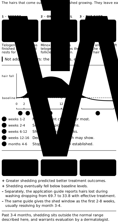
Why minoxidil makes hair fall out first, and how long the shed runs for
Two parts. First, the mechanism, drawn as one follicle in three states. Before: the follicle is shallow and the hair in it has finished growing, resting for two to four months in telogen. On minoxidil: the drug shortens telogen and moves the follicle back into anagen, so the follicle deepens and a new shaft forms beneath the old finished hair. The shed: the new shaft comes up through the scalp and pushes the old, weak telogen hair out. These are not additional hairs; they are the same hairs, leaving earlier. Second, a timeline of hair fall over the weeks and months after starting. The vertical axis has no scale, because no source gives one; the curve shows shape only. It sits at baseline through weeks one to two, when most notice no significant changes; begins rising in weeks two to four, when shedding typically begins; peaks across weeks six to twelve; falls through weeks twelve to sixteen as shedding decreases and new growth may become visible; and is flat again through months four to six, when shedding has stopped and new growth is established. The curve ends just below the baseline it started from, because shedding is reported to have eventually fallen below baseline levels. Separately, the application guide reports hairs lost during washing dropping from an average of 69.7 to 33.8 with effective treatment. Shedding is reported in seventeen to fifty-five percent of users, with an average duration of one and a half to two months, and most users see improvement by weeks twelve to sixteen. Greater shedding predicted better treatment outcomes. The same guide gives the shed window as the first two to eight weeks, usually resolving by month three to four. Past three to four months, shedding sits outside the normal range described here, and warrants evaluation by a dermatologist.
The hairs that come out had already finished growing. They leave early.
What minoxidil does to a resting follicle
1 · BEFORE
Telogen: the hair has
finished growing and
rests for 2-4 months.
2 · ON MINOXIDIL
Minoxidil shortens
telogen and moves the
follicle into anagen.
3 · THE SHED
The new anagen shaft
pushes the old, weak
telogen hair out.
Not additional hairs: the same hairs, earlier.
The shedding window, in weeks after starting
hair fall
baseline
0
2
4
6
8
12
16
1
2
3
4
5
1
weeks 1-2
No significant changes for most.
2
weeks 2-4
Shedding typically begins.
3
weeks 6-12
Shedding typically peaks.
4
weeks 12-16
Decreases; new growth may show.
5
months 4-6
Stopped; new growth established.
What the studies report
17-55%
of users are reported
to shed
1.5 to 2
months, the average
duration of the shed
12-16
weeks: most see
improvement by here
+ Greater shedding predicted better treatment outcomes.+ Shedding eventually fell below baseline levels.– Separately, the application guide reports hairs lost duringwashing dropping from 69.7 to 33.8 with effective treatment.
– The same guide gives the shed window as the first 2-8 weeks,usually resolving by month 3-4.
Past 3-4 months, shedding sits outside the normal range
described here, and warrants evaluation by a dermatologist.
Minoxidil shortens telogen and moves resting follicles back into growth, so the old club hairs — which had already finished growing — are pushed out as the new shafts come up. The curve shows the shape reported after starting: a rise, a peak, a fall, and a settling below where it began. The vertical axis carries no scale because no source states one. Appears in How to Stop Minoxidil Shedding Phase: Complete Guide (2026) · Minoxidil 5% Complete Guide: How to Apply for Best Results .
Use this figure on your own site
Free to use anywhere, including commercially, if you keep a visible link back to this page. It is drawn from the sources cited beneath it and follows your reader's light or dark theme.
Open the SVG directly · All figures
The Norwood scale, stages 1 to 7 and the Type A variant#
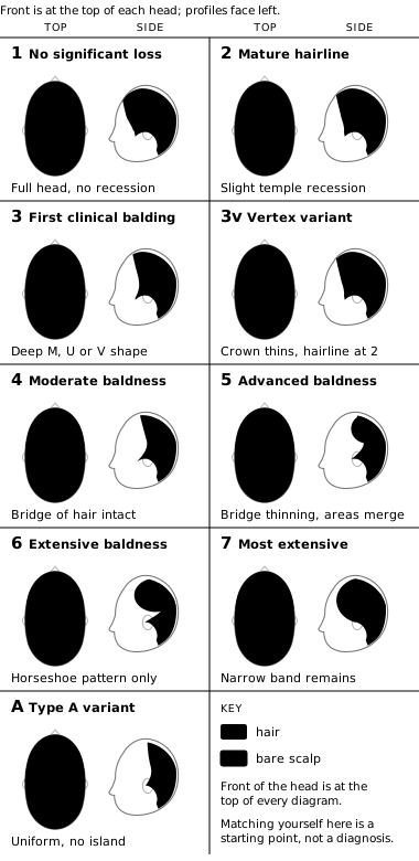
The Norwood scale: stages 1 to 7, the stage 3 vertex variant, and the Type A variant
Nine simplified head diagrams. Each stage is shown twice, once from above with the front of the head at the top and once in profile facing left. Shaded areas are hair; pale areas are bare scalp. Stage 1, No significant loss: The baseline the other stages are measured against. Stage 2, Mature hairline: Usually normal maturation rather than balding. Stage 3, First clinical balding: The first stage the scale counts as balding. Stage 3v, Vertex variant: Crown thinning behind a Stage 2 hairline. Stage 4, Moderate baldness: A bridge of hair still separates front from crown. Stage 5, Advanced baldness: The two bald areas are beginning to merge. Stage 6, Extensive baldness: Front and crown are one field; a horseshoe remains. Stage 7, Most extensive: Only a narrow band around the sides and back. Stage A, Type A variant: Recedes evenly, and shows no separate crown loss. Matching yourself against the scale is a starting point, not a diagnosis.
Front is at the top of each head; profiles face left.
TOP
SIDE
TOP
SIDE
1
No significant loss
Full head, no recession
2
Mature hairline
Slight temple recession
3
First clinical balding
Deep M, U or V shape
3v
Vertex variant
Crown thins, hairline at 2
4
Moderate baldness
Bridge of hair intact
5
Advanced baldness
Bridge thinning, areas merge
6
Extensive baldness
Horseshoe pattern only
7
Most extensive
Narrow band remains
A
Type A variant
Uniform, no island
KEY
hair
bare scalp
Front of the head is at the
top of every diagram.
Matching yourself here is a
starting point, not a diagnosis.
Each stage is drawn twice: the top of the head with the front at the top, and the profile facing left. Shaded areas are hair, pale areas are bare scalp. Stage 2 is usually a mature hairline rather than balding; Stage 3 is the first stage the scale counts as clinical balding. The Type A variant recedes uniformly from front to back instead of leaving an island of hair in the middle, and shows no separate crown loss. Appears in The Norwood Scale Explained: Find Your Stage of Hair Loss (2026) .
Use this figure on your own site
Free to use anywhere, including commercially, if you keep a visible link back to this page. It is drawn from the sources cited beneath it and follows your reader's light or dark theme.
Open the SVG directly · All figures
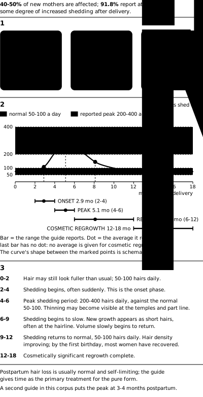
The postpartum shedding curve#
The postpartum shedding curve: what pregnancy holds, what delivery releases, and when density returns
A three-part plate about hair shedding after childbirth. Part one, three panels of a follicle in section. In pregnancy, high estrogen extends the growth phase, so every follicle is drawn deep and attached and fewer hairs fall out. At delivery, estrogen drops rapidly and the held hairs shift to the resting phase together: the shafts are still attached but clubbed at the root. About 2 to 3 months later they shed at once, roughly 9 months of hair that was held back; the follicles they leave are drawn intact, not damaged. Part two, a curve of daily shedding against months after delivery. A lower band marks normal shedding of 50 to 100 hairs a day and an upper band the reported peak of 200 to 400 a day. The curve sits in the normal band for the first two months, climbs steeply through months 3 and 4, peaks around month 5, and falls back into the normal band by about month 12. Beneath it, four bars on the same month scale: onset averages 2.9 months with a reported range of 2 to 4; the peak averages 5.1 months, range 4 to 6; shedding resolves at an average of 8.1 months, range 6 to 12; and cosmetically significant regrowth takes 12 to 18 months, a range for which no average is given, so that bar carries no dot. The shape of the curve between the marked points is schematic. Part three, the month-by-month description: months 0 to 2, hair may still look fuller than usual at 50 to 100 hairs daily; months 2 to 4, shedding begins, often suddenly, the onset phase; months 4 to 6, peak shedding of 200 to 400 hairs daily, with thinning possibly visible at the temples and part line; months 6 to 9, shedding slows and new growth appears as short hairs at the hairline; months 9 to 12, shedding returns to normal and density improves, with most women recovered by the first birthday; months 12 to 18, cosmetically significant regrowth complete. Postpartum hair loss affects 40 to 50 percent of new mothers, with 91.8 percent reporting at least some increased shedding. It is usually normal and self-limiting, and the guide gives time as the primary treatment. A second guide in this corpus puts the peak earlier, at 3 to 4 months postpartum.
40-50% of new mothers are affected; 91.8% report at leastsome degree of increased shedding after delivery.
1 PREGNANCY HOLDS, DELIVERY RELEASES
PREGNANCY
Estrogen extends the
growth phase. Fewer
hairs fall out.
DELIVERY
Estrogen drops fast.
Held hairs shift to
telogen together.
2-3 MONTHS LATER
They shed at once:
about 9 months of
hair held back.
2 THE SHEDDING CURVEhairs shed daily
normal 50-100 a day
reported peak 200-400 a day
50 100 200 400
0 2 4 6 8 10 12 14 16 18
months after delivery
ONSET 2.9 mo (2-4) PEAK 5.1 mo (4-6) RESOLVES 8.1 mo (6-12) COSMETIC REGROWTH 12-18 mo
Bar = the range the guide reports. Dot = the average it reports. The
last bar has no dot: no average is given for cosmetic regrowth.
The curve's shape between the marked points is schematic.
3 MONTH BY MONTH, AS THE GUIDE DESCRIBES IT0-2 Hair may still look fuller than usual; 50-100 hairs daily. 2-4 Shedding begins, often suddenly. This is the onset phase. 4-6 Peak shedding period: 200-400 hairs daily, against the normal 50-100. Thinning may become visible at the temples and part line. 6-9 Shedding begins to slow. New growth appears as short hairs, often at the hairline. Volume slowly begins to return. 9-12 Shedding returns to normal, 50-100 hairs daily. Hair density improving; by the first birthday, most women have recovered. 12-18 Cosmetically significant regrowth complete.
Pregnancy holds hairs in the growth phase; after delivery they shift to resting together and shed a few months later, all at once. The guide reports shedding beginning at 2.9 months on average, peaking at 5.1 and resolving at 8.1, with cosmetically significant regrowth by 12-18 months. The bars are the ranges reported, the dots the averages; the curve between the marked points is schematic. The guide reports the course as usually self-limiting. Appears in Hair Loss After Pregnancy: Recovery Guide .
Use this figure on your own site
Free to use anywhere, including commercially, if you keep a visible link back to this page. It is drawn from the sources cited beneath it and follows your reader's light or dark theme.
Open the SVG directly · All figures
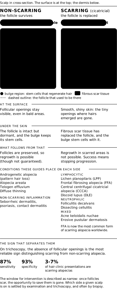
Scarring and non-scarring hair loss, in cross-section#
Scarring and non-scarring alopecia compared in scalp cross-section
Two cross-sections of scalp side by side, the skin surface at the top and the dermis below. Left, non-scarring alopecia: the skin line is broken by three follicular openings, and beneath each one a whole follicle runs down to its bulb, with the bulge region marked on its side as the site of the stem cells that regenerate hair. One follicle carries a full hair, one a fine short hair, and the middle one none at all, yet it is intact: the follicle is dormant, not dead, so regrowth remains possible though it is not guaranteed. Right, scarring or cicatricial alopecia: the skin line runs unbroken, with no openings anywhere, and where each follicle used to be there is a field of fibrous scar tissue, the lost follicle shown only as a dashed ghost and the bulge marker struck through. Nothing emerges from the surface. Regrowth in scarred areas is not possible, and success means stopping progression. Conditions these guides place on the non-scarring side: androgenetic alopecia or pattern hair loss, alopecia areata, telogen effluvium, diffuse thinning, and the non-scarring inflammatory conditions seborrheic dermatitis, psoriasis and contact dermatitis. On the scarring side, grouped as the North American Hair Research Society classifies them: lymphocytic, being lichen planopilaris, frontal fibrosing alopecia, central centrifugal cicatricial alopecia and discoid lupus; neutrophilic, being folliculitis decalvans and dissecting cellulitis; and mixed, being acne keloidalis nuchae and erosive pustular dermatosis. Frontal fibrosing alopecia is now the most common form of scarring alopecia worldwide. On trichoscopy, absence of the follicular openings is the most reliable sign distinguishing the two, with 87 percent sensitivity and 93 percent specificity; scarring alopecias represent 3 to 7 percent of hair clinic presentations. The window for intervention is described as narrow: once follicles scar, the opportunity to save them is gone.
Scalp in cross-section. The surface is at the top; the dermis below.
NON-SCARRING
the follicle survives
Regrowth remains possible
SCARRING(cicatricial)
the follicle is replaced
The loss is permanent
opening (ostium) no opening bulge region: stem cells that regenerate hair
fibrous scar tissue
dashed outline: the follicle that used to be there
AT THE SURFACE Follicular openings stay
visible, even in bald areas.
Smooth, shiny skin: the tiny
openings where hairs
emerged are gone.
UNDER THE SKIN The follicle is intact but
dormant, and the bulge keeps
its stem cells.
Fibrous scar tissue has
replaced the follicle, and the
bulge stem cells with it.
WHAT FOLLOWS FROM THAT Follicles are preserved, so
regrowth is possible
(though not guaranteed).
Regrowth in scarred areas is
not possible. Success means
stopping progression.
CONDITIONS THESE GUIDES PLACE ON EACH SIDE Androgenetic alopecia
(pattern hair loss)
Alopecia areata
Telogen effluvium
Diffuse thinning
NON-SCARRING INFLAMMATION
Seborrheic dermatitis,
psoriasis, contact dermatitis
LYMPHOCYTIC
Lichen planopilaris (LPP)
Frontal fibrosing alopecia (FFA)
Central centrifugal cicatricial
alopecia (CCCA)
Discoid lupus (DLE)
NEUTROPHILIC
Folliculitis decalvans
Dissecting cellulitis
MIXED
Acne keloidalis nuchae
Erosive pustular dermatosis
FFA is now the most common form
of scarring alopecia worldwide.
THE SIGN THAT SEPARATES THEM
On trichoscopy, the absence of follicular openings is the most
reliable sign distinguishing scarring from non-scarring alopecia.
87%
sensitivity
93%
specificity
3-7%
of hair clinic presentations are
scarring alopecias
The window for intervention is described as narrow: once follicles
scar, the opportunity to save them is gone. Which side a given scalp
is on is settled by examination and trichoscopy, and often by biopsy.
Two scalp cross-sections. Where the alopecia is non-scarring the follicle survives and its opening at the surface stays visible, so regrowth remains possible; where it is scarring (cicatricial) fibrous tissue has replaced the follicle, the opening is gone, and that loss is permanent. Which of the two a scalp shows is settled by examination and trichoscopy, not from a diagram. Appears in Scarring Alopecia Types and Management: Complete Guide · Frontal Fibrosing Alopecia: Complete Guide to Symptoms, Causes & Treatment (2026) .
Use this figure on your own site
Free to use anywhere, including commercially, if you keep a visible link back to this page. It is drawn from the sources cited beneath it and follows your reader's light or dark theme.
Open the SVG directly · All figures
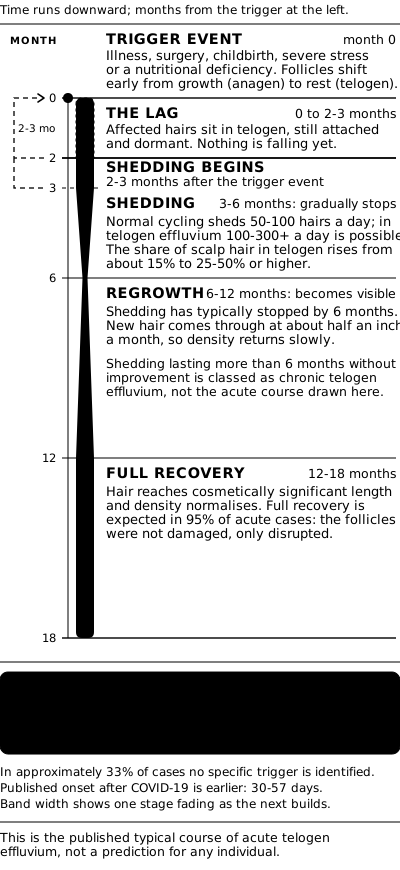
Why the shedding starts months after the thing that caused it#
The published course of acute telogen effluvium, from the trigger to full recovery
A timeline read downward on a single linear scale of months from a triggering event. Month 0, the trigger: illness, surgery, childbirth, severe stress or a nutritional deficiency, which makes follicles shift early from growth into rest. Nothing is visible yet. From month 0 to months 2-3, the lag: the affected hairs sit in telogen, still attached and dormant, and nothing is falling. An arrow runs from the shedding line back up to the trigger, marking a gap of 2-3 months. At 2-3 months, shedding begins. From then until months 3-6 the shedding band tapers as shedding gradually stops; normal cycling sheds 50 to 100 hairs a day, while in telogen effluvium 100 to 300 or more a day is possible, and the share of scalp hair in telogen rises from about 15 percent to 25-50 percent or higher. Shedding has typically stopped by 6 months; shedding lasting more than 6 months without improvement is the chronic form rather than the acute course drawn here. From 6 to 12 months regrowth becomes visible, with new hair growing about half an inch a month. From 12 to 18 months hair reaches cosmetically significant length and density normalises; full recovery is expected in 95 percent of acute cases, because the follicles were not damaged, only disrupted. The reason the cause is hard to find: telogen lasts about 3-4 months, so a hair pushed into it early falls on that clock and the shedding appears 2-3 months after the event that set it off. In approximately 33 percent of cases no specific trigger is identified, and published onset after COVID-19 is earlier, at 30 to 57 days. The width of each band shows one stage fading as the next builds; it is not a count of hairs. This is the published typical course of acute telogen effluvium, not a prediction for any individual.
Time runs downward; months from the trigger at the left.
MONTH
0
2
3
6
12
18
2-3 mo
TRIGGER EVENT month 0
Illness, surgery, childbirth, severe stress or a nutritional deficiency. Follicles shift early from growth (anagen) to rest (telogen).
THE LAG 0 to 2-3 months
Affected hairs sit in telogen, still attached and dormant. Nothing is falling yet.
SHEDDING BEGINS
2-3 months after the trigger event
SHEDDING 3-6 months: gradually stops
Normal cycling sheds 50-100 hairs a day; in telogen effluvium 100-300+ a day is possible. The share of scalp hair in telogen rises from about 15% to 25-50% or higher.
REGROWTH 6-12 months: becomes visible
Shedding has typically stopped by 6 months. New hair comes through at about half an inch a month, so density returns slowly.
Shedding lasting more than 6 months without improvement is classed as chronic telogen effluvium, not the acute course drawn here.
FULL RECOVERY 12-18 months
Hair reaches cosmetically significant length and density normalises. Full recovery is expected in 95% of acute cases: the follicles were not damaged, only disrupted.
WHY THE CAUSE IS HARD TO FIND
Telogen lasts about 3-4 months, so a hair pushed into it early falls on that clock. The shedding shows up 2-3 months after the event that set it off, not in the week it starts.
In approximately 33% of cases no specific trigger is identified. Published onset after COVID-19 is earlier: 30-57 days. Band width shows one stage fading as the next builds.
This is the published typical course of acute telogen effluvium, not a prediction for any individual.
The published course of acute telogen effluvium on a single linear month axis: the trigger, the lag while the affected hairs sit in telogen, the shedding window, then regrowth and full cosmetic recovery. The arrow marks the gap that makes the cause hard to find. This is the typical course reported in the literature, not a prediction for any individual. Appears in Telogen Effluvium: Causes and Recovery Timeline .
Use this figure on your own site
Free to use anywhere, including commercially, if you keep a visible link back to this page. It is drawn from the sources cited beneath it and follows your reader's light or dark theme.
Open the SVG directly · All figures